Hand and Wrist Surgery

Those with severe musculoskeletal and arthritis-related conditions in their hand and wrist may wish to have surgery, especially if other treatments (such as medication and therapy) have not worked. As specialists in the area of the hand and wrist, our doctors perform the following surgeries:

Jump to: Carpal Tunnel Syndrome | DeQuervain’s Tenosynovitis | Hand Arthritis | Dupuytren’s Contracture | Trigger Finger | Fractures of the Hand and Wrist | TFCC and DRUJ Injuries

Contact us today for a consultation »

hand and wrist surgery california

Carpal Tunnel Syndrome

Patients often present to the office concerned about having “carpal tunnel” because they have pain in the hand. Carpal tunnel generally does not cause pain but rather numbness and tingling. It is a compression phenomenon on the median nerve at the palm (at the wrist crease). It is aggravated by wrist flexion and extension which is why it often presents with numbness and tingling at night (as people often sleep with the wrist(s) flexed or extended).

The median nerve provides sensation to the palmar side of the hand, in the thumb, index, middle and half of the ring finger. It also provides motor function to the thenar muscles (the thumb muscles in the palm). The diagnosis of carpal tunnel syndrome is made with a history of numbness and tingling in the fingers, with physical exam (the Tinel’s, Phalen’s and compression tests are three commonly used tests to assess for carpal tunnel syndrome) and a nerve study can also provide the doctor information as to the presence and severity of carpal tunnel.

The first line of treatment for carpal tunnel is activity modification. Namely, avoiding activities that aggravate the condition (wrist flexion and extension, activities that cause vibration of the hand, for instance). A wrist brace, keeping the wrist immobilized in neutral position can help prevent the symptoms with activities of daily living and when sleeping at night.

If bracing does not work, a cortisone injection into the carpal tunnel can settle down the inflammation within the carpal tunnel and can help with the symptoms for days, weeks and potentially for the long-term.

For patients who fail to improve with conservative management, the surgical treatment for carpal tunnel is a carpal tunnel release. This is performed either by making an incision in the palm to release the transverse carpal ligament (the ligament that is compressing the nerve, causing the symptoms) or by releasing the ligament endoscopically (with a camera). There are advantages and disadvantages to both procedures but the literature shows that the outcomes of the procedures are similar.

The procedure takes approximately 15 minutes and it is generally performed in an operating room under local anesthesia or sedation. The wrist is immobilized after surgery for 7-10 days. Thereafter, early motion is encouraged and patients are arranged for hand therapy, if necessary. Full recovery, on average, is 3 months.

mommy's thumb treatment

DeQuervain’s Tenosynovitis

DeQuervain’s Tenosynovitis (also known as “Mommy’s Thumb”) is an inflammatory condition of two tendons in the wrist. The two tendons (the APL and EPB tendons) run underneath a sheath (retinaculum) that can get inflamed from overuse. The most common cause of this condition is excessive thumb abduction (moving the thumb away from the index finger). This action is common when opening jars, for instance, and when picking things up (such as a child). This malady is common in parents with young children because they are often doing a lot of opening of jars and of picking up of their children.

DeQuervain’s presents as a sharp, oftentimes debilitating, pain in the hand and wrist, just below the base of the thumb. It can cause swelling in the region and the sheath overlying the tendons can get so inflamed that it thickens and it can become more visible than the unaffected side. It can also cause clicking in extreme cases, as the tendons can thicken from the inflammation and this can cause clicking of the tendons under the sheath with thumb abduction.

The first line of treatment for DeQuervain’s is activity modification. Avoid the activities that aggravate the condition, if possible. Wearing a thumb spica splint (a splint that immobilizes the wrist and the thumb) can help as well. A cortisone injection is often very helpful both in the short and long-term for the pain but without activity modification, the symptoms may recur. Generally, if the patient does not improve with activity modification, bracing and a cortisone injection, the definitive treatment for DeQuervain’s is decompression of the two tendons by splitting the sheath overlying the tendons (a “first dorsal compartment release”). This procedure takes about 15 minutes and is typically performed in an operating room under local anesthesia or sedation. The wrist is immobilized for about a week after surgery. Thereafter, the patient is encouraged to get the wrist moving and is arranged for hand therapy, if necessary. Full recovery is generally 3 months.

Contact us today for a consultation »

Hand Arthritis

Arthritis in the hand is very common, particularly in patients over 50 years of age. Arthritis – in any joint in the body – is degeneration of the cartilage. “Arth” (joint) “itis” (inflammation) can have numerous causes, including trauma to a joint, infection, and most commonly, age-related wear and tear of the joint cartilage (“degenerative arthritis”). This is in contrast to Rheumatoid Arthritis which is an auto-immune condition in which the body’s own immune system attacks joint cartilage, causing degeneration of the cartilage. Arthritis caused by Rheumatoid Arthritis has become less common in the last decade with the advent of medications which can prevent the body from attacking its own joints.

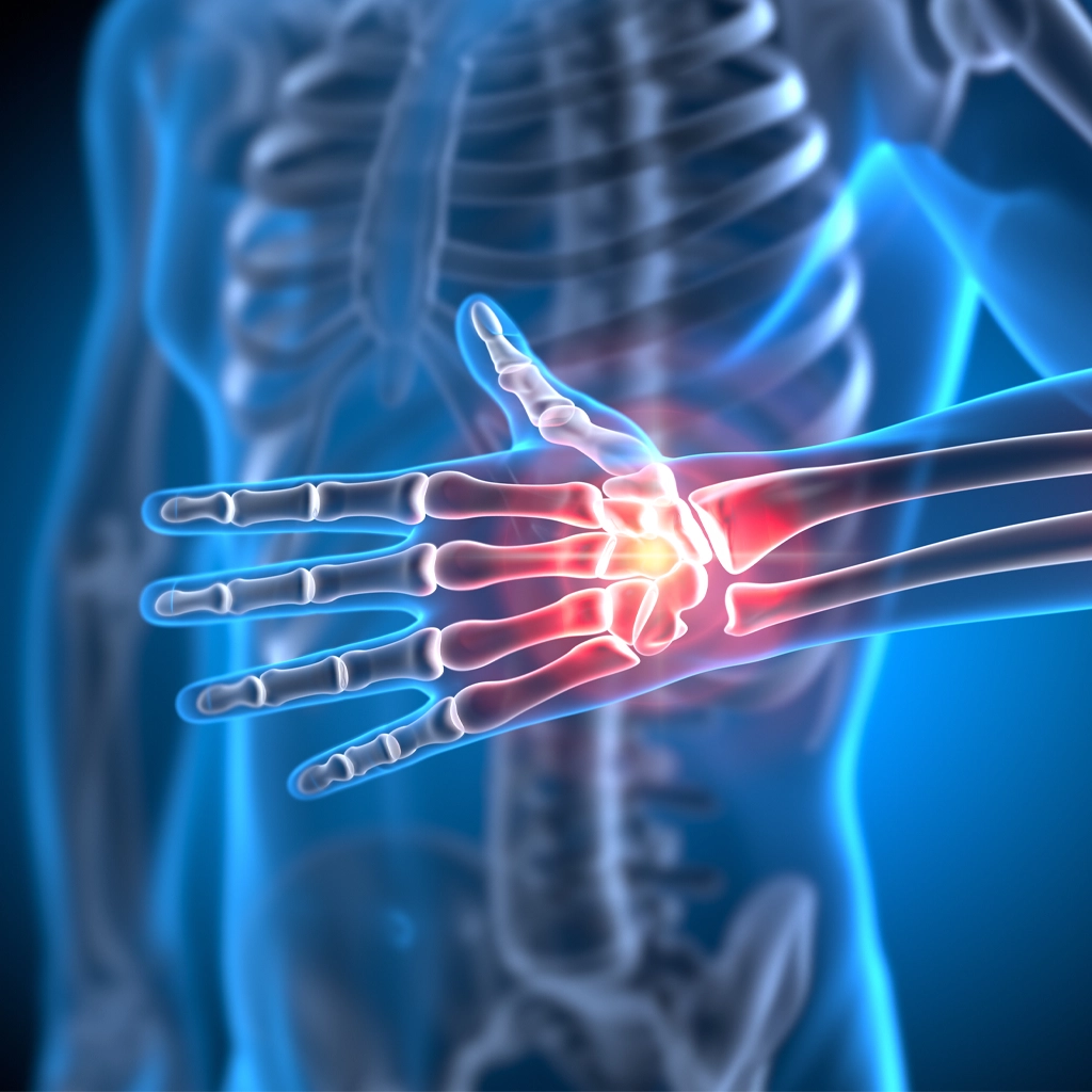
Arthritis of the hand can affect any joint of the hand or wrist. The joint that most commonly becomes arthritic in the hand is the joint at the base of the thumb (the carpometacarpal joint or “CMC” joint). Arthritis causes pain, stiffness and with advanced degeneration, deformity of the joint.

The first line of treatment for arthritis (of any joint) is activity modification. Avoid activities that aggravate the pain in the joint. Non-steroidal anti-inflammatory medications are very commonly used to treat pain associated with arthritis. Unfortunately, there is no way to reverse the changes that occur to the joint when it becomes arthritic. Cartilage is very durable but once it deteriorates, there remains no method by which the cartilage can be restored to new.

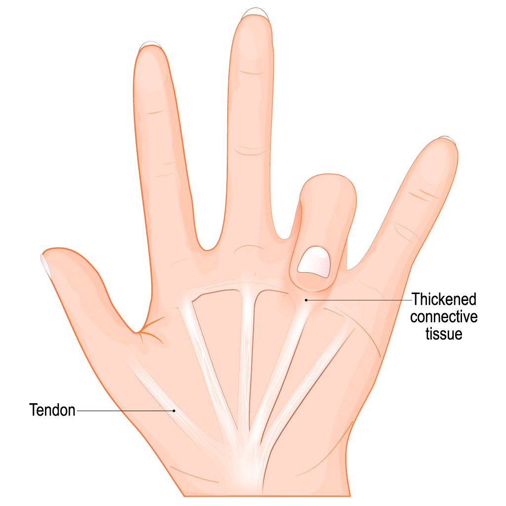
A cortisone injection can provide temporary (weeks to months) of relief from the pain associated with arthritis. A patient can receive a cortisone injection two or three times per year. If given too often, cortisone can cause permanent damage to the tendons and ligaments surrounding the joint. This is why doctors are judicious in how often they administer cortisone.

For thumb CMC joint arthritis, a splint can help alleviate the symptoms. If a patient fails to improve with splinting and an occasional cortisone injection, surgery can provide long-term relief. There are several surgical techniques for thumb CMC arthritis. A common technique is removal of the trapezium bone (the carpal bone that articulates with the base of the thumb metacarpal at the CMC joint). A soft tissue spacer is sometimes place in the void left by the removed bone, to prevent subsidence (settling) of the thumb. Generally, the thumb is immobilized for 4-6 weeks after surgery. Thereafter, the patient is placed in a removable splint, arranged for hand therapy and encouraged to get the thumb moving. Full recovery from the procedure is, on average, 6 months.

For other joints in the hand, splinting is generally not particularly helpful for arthritis. Cortisone injections can be helpful but there is often not a lot of space in the joint into which the doctor can place cortisone. If a patient fails conservative management for arthritis in the hand, the surgical options include fusing the arthritic joint or replacing the joint with a rubber (or metal) joint. The advantage of a fusion is that it is a one and done surgery; after fusing the joint (removing the remaining cartilage and compressing the bones together, to heal, like a fracture), the pain should be gone forever. The drawback is that the patient will lose motion at that joint.

The advantage of a joint replacement is that it can preserve motion of the joint. The disadvantage is that it is like a tire. It will wear out with time and will either require a revision joint replacement or a conversion to a fusion. Thus, if a patient opts for a finger joint replacement, then the patient should be made aware of the fact that a second surgery may be necessary in the future, to either replace the joint replacement or to fuse the joint.

Joint surgeries for hand arthritis are performed under general anesthesia. Surgical time is, on average, about an hour per joint. Full recovery is 3-6 months.

Dupuytren’s Contracture

Dupuytren’s Contracture is the contracture of the palmar fascia (a thick tissue underneath the palmar skin). To this day, we are not sure why people develop Dupuytren’s Contracture but there are risk factors. The most common risk factor is being of Northern European descent. Although we do not fully understand why patients develop it, we do understand what is occurring to the tissue, to cause the deformity.

Dupuytren’s Disease treatment

Patients with Dupuytren’s Contracture oftentimes present complaining of deformity in the palm. There can be pitting of the skin within the palm, as well as firm nodules in the palm. The small and ring finger are most commonly affected although all four fingers and the thumb can be affected. If the patient has pitting in the palm and/or nodules but no flexion contracture of the fingers, this is called “Dupuytren’s Disease.” Once the diseased tissue extends into the fingers, causing a fixed flexion contracture of the finger, this is “Dupuytren’s Contracture.” The disease moves from the palm into the finger, first at the MCP (metacarpophalangeal) joint and sometimes it can extend all the way to the PIP (proximal interphalangeal) joint. As a result of the contracture, patients can lose the ability to fully extend one or more fingers. This can affect one’s ability to perform basic activities of daily living, such as putting your hand into your pocket.

The treatment for Dupuytrens Disease (no contracture of the fingers) is usually observation. The tabletop test is a simple test for patients to perform. If you a patient can lay the hand flat on a table, it is generally best to observe. On occasion, a patient will find the diseased tissue within the palm painful and will opt for surgical excision of the tissue, even if there is no contracture. This is done under general anesthesia and can be curative but the diseased tissue can recur or can subsequently affect another finger (or the other hand).

Once a patient develops any degree of contracture at a finger MCP joint, the recommendation is to intervene in some way. The reason for this is, the worse the contracture gets, the harder it can be to treat and the fewer options available to treat the condition.

A commonly used technique that became available circa 2010 is Xiaflex. Xiaflex is an enzyme (collagenase) that can break up the tissue that causes Dupuytren’s Contracture. It does not eradicate the tissue, meaning the tissue remains underneath the skin, but the enzyme can break up the tissue, to eradicate the contracture itself. The protocol for this involves two office visits: In the first visit, the doctor will inject the Xiaflex into the “cord” (the diseased tissue causing the contracture). The second visit (one or two days later) involves manipulating the finger (gently) in the office (after injecting local anesthetic into the finger) in order to break up the diseased tissue.

The most common side effects of a Xiaflex injection are swelling, tenderness, pain, bruising and skin tears in the region where the cord is broken. The skin tears are treated with wound care until the wounds heal.

Even if Xiaflex is successful in breaking up the diseased tissue, the disease can recur. If it does, Xiaflex can be used again. It usually takes months to years for a recurrence, not days to weeks.

Another technique that is less commonly used for Dupuytren’s contracture is needle aponeurotomy. This technique is performed in the office under local anesthesia. The doctor uses a needle to cut the diseased tissue with small sweeps of the needle. The advantage of this is it is minimally invasive. The risk of this is that the doctor could injure a nerve, blood vessel or tendon when swiping with the needle. Even if the procedure is successful, the contracture can recur.

Surgery is the gold standard for removing Dupuytren’s Contracture. This is done under general anesthesia and can take an hour or two, depending on the severity of the contracture and the number of fingers affected. The advantage of surgery is that it actually removes the tissue and can provide a long-term solution. The disadvantage of surgery is it is painful and the recovery is 3-6 months. Even with surgery, the disease can recur.

Contact us today for a consultation »

Trigger Finger

Trigger finger is one of the most common hand conditions. The reason it is called a “trigger finger” is because the finger can lock in flexion and it looks like someone pulling a trigger.

Trigger finger is caused by inflammation of the A1 pulley – a sheath overlying the flexor tendons in the palm. There are five pulleys in each finger. The A1 pulley is located in the palm, at the level of the metacarpal head (the “big knuckle”). When a patient has a trigger finger, there does not have to have triggering. In fact, a lot of patients with trigger fingers never have triggering. Rather, the patient presents with a sharp pain in the palm. The diagnosis is made with history and physical exam. X-rays and/or MRI are not necessary to make the diagnosis. Patients are tender at the A1 pulley of the finger. On occasion, the patient will be able to demonstrate triggering of the finger. On rare occasion, a patient’s finger will be stuck in a flexed position because of the severity of the trigger finger.

The most common treatment for a trigger finger is a cortisone injection into the A1 pulley. This is oftentimes curative. If a patient’s symptoms persist after one cortisone injection, then the doctor and the patient can discuss either trying one more injection (at least one month after the first injection) or proceeding with surgery. Surgery for a trigger finger can be done under local anesthesia or sedation. It in involves making a small incision over the A1 pulley and splitting the pulley. Fortunately, we do not need our A1 pulley to have completely normal finger function. The procedure takes about 10 to 15 minutes. The hand is splinted for about 10 days after surgery at which time the sutures are removed and the patient is arranged for hand therapy, if needed. Full recovery is, on average, 3 months.

hand fracture treatment

Fractures of the Hand and Wrist

Fractures of the hand and wrist are very common and they occur in all age groups. In general, the treatment of fractures depends on two main factors: The degree of displacement of the fracture (how much has it moved from where it is supposed to be?) and whether or not the fracture involves a joint. Non-displaced fractures, on the whole, can be treated with 4-6 weeks of immobilization (splint or a cast). Fractures that involve a joint (intra-articular fractures) can also be treated with immobilization, if the fracture is non-displaced or minimally displaced (less than 2 mm). If a fracture has significant displacement and/or it has significant intra-articular displacement, then it will require some sort of intervention.

Possible interventions for displaced fractures include manipulation of the fracture (in the emergency room, in the office, or in an operating room) to improve the fracture alignment. The fracture is then held in place with a splint or a cast.

If a fracture of the hand or wrist is not amenable to casting or splinting (with or without manipulation), then the fracture may require fixation with a plate and screws (in a wrist fracture, for instance) or with a single screw (for a scaphoid fracture). The purpose of the plates and screws is to hold the fractured bone in an anatomic position so it will heal correctly. If a displaced fracture is not “reduced” back to its normal position, so it can heal correctly, it may heal, but it will do so in a way which can lead to chronic pain, deformity and limited functionality.

Two of the most common fractures of the upper extremity are a distal radius fracture (wrist fracture) and scaphoid fracture. Both of these fractures are common after a fall onto an outstretched wrist. Distal radius fractures are common in children, adolescents and adults alike. Scaphoid fractures are most common in active individuals in their 20s or 30s. Scaphoid fractures are common in skateboarders and snowboarders, for instance.

The scaphoid is a peanut-shaped bone within the carpus (the small bones of the wrist). It is located between the thumb and the end of the radius bone. It typically causes pain in the anatomic snuffbox (the soft spot between the thumb and the radius). It is not uncommon for scaphoid fractures to be missed on X-rays because they can be hard to see. If a patient has a history and physical exam concerning for a scaphoid fracture, an MRI is generally ordered, as it will show a fracture that may not be evident on X-rays. The treatment of scaphoid fractures depends on the degree of displacement of the fracture and the location of the fracture within the scaphoid. The scaphoid has poor blood supply and as a result, it heals more slowly than most bones. Also, the more proximal the fracture (the area of the scaphoid closer to the radius), the more likely the fracture will not heal without fixation. Whether a scaphoid fracture requires surgery or not, on average, a scaphoid heals in 8-10 weeks. If the fracture is amenable to conservative management, a patient can expect at least 6-8 weeks of immobilization of the wrist and thumb, to allow the fracture ample time to heal.

All fractures should first be evaluated with X-rays. Sometimes, depending on the type of fracture, an MRI or CT may be required but on the whole, X-rays are sufficient for the evaluation of hand and wrist fractures.

Many distal radius fractures can be treated conservatively, with casting or splinting, as long as the fracture is not significantly displaced. If the fracture is displaced, then there are different techniques for fixing a distal radius fracture. In children, this generally involves closed reduction and casting under anesthesia. In adults, this usually involves fixation of the fracture with a plate and screws.

Recovery from hand and wrist fractures, no matter the treatment method, is variable, depending on a patient’s age and the severity of the fracture.

TFCC and DRUJ Injuries

The TFCC (triangular fibrocartilage complex) and DRUJ (distal radial-ulnar joint) are two structures that commonly cause pain on the ulnar side of the wrist (the small finger side of the wrist). There are two bones that make up the wrist, the radius and ulna. The radius and ulna are stabilized to one another with both the DRUJ and TFCC. Both of these structures can be injured, commonly with a fall onto an outstretched wrist. Patients typically present with pain in the ulnar aspect of the wrist, in the region of the TFCC and/or DRUJ. The evaluation of these injuries involves a history, physical exam and X-rays. Depending on the findings, an MRI arthrogram (dye is injected into the wrist joint) may be necessary in order to diagnose the injury. TFCC injuries are far more common than DRUJ injuries. A DRUJ injury can cause instability of the joint between the radius and ulna. This often occurs in conjunction with a wrist or forearm fracture. TFCC injuries can occur with less significant trauma. For instance, it is not uncommon for patients to injury the TFCC playing tennis, doing push-ups or yoga.

If the DRUJ is unstable, it requires immobilization and oftentimes surgical fixation, most commonly with pinning of the joint for 6 weeks, to allow the DRUJ ligaments to heal.

The TFCC has a couple of purposes. It helps stabilize the radius and ulna and it acts as a bumper between the head of the ulna and the triquetrum (a carpal bone). Without the TFCC, the head of the ulna and the triquetrum would hit each other with certain wrist motions.
There are numerous types of TFCC injuries. In general, the first line of treatment for most TFCC injuries is a period of immobilization (4-6 weeks), activity modification, non-steroidal anti-inflammatory medications, and sometimes patients opt for a cortisone injection in to the TFCC region, for pain relief. Hand therapy can be a useful adjunct for TFCC injuries, as well.

For TFCC injuries that are refractory to conservative management, the most common surgical option is arthroscopic surgery of the wrist. This is performed under general anesthesia. It requires small incisions on the back of the wrist. The doctor assesses the wrist joint, including the TFCC, using a small camera. Depending on the type of TFCC tear, the surgeon will either clean out the TFCC tear (arthroscopic debridement) or perform an arthroscopic TFCC repair. Sometimes, a TFCC tear requires an “open” (non-arthroscopic) repair, whereby the surgeon makes an incision on the back of the wrist, looks at the TFCC under direct visualization and repairs the TFCC (typically with suture).

Recovery from a TFCC surgery depends on whether the TFCC was repaired or “cleaned out.” A repair typically requires one month of immobilization. A clean out generally does not require immobilization for more than 7-10 days after surgery. Hand therapy is frequently required for 12 weeks after surgery and full recovery from TFCC surgery is 3-6 months, on average.

If you are interested in learning more about Golden State Bone and Joint services and hand and wrist condition treatments, contact us today!

Skip to content