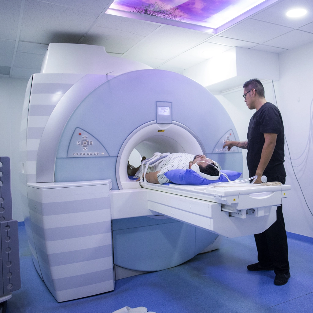
With the opening of our new surgery center, the Siemens MAGNETOM Aera high-field strength MRI is available to assist in diagnosing a wide variety of orthopaedic conditions. In addition to greater convenience for patients who won’t have to visit another imaging center for their diagnostic testing, our new MRI offers faster scan times with fewer re-takes required.
This MRI system is specially made for orthopaedic use and is capable of obtaining exceptionally high-quality images that can be reconstructed into 3D images to assist in precise surgical planning.
The two types of electrodiagnostic testing we offer are electromyography and nerve condition studies. They measure electrical activity produced by the nerves and muscles in the body, and can help determine if you have an injury that is impacting nerve or muscle function. It is commonly used to evaluate conditions such as peripheral neuropathy, carpal tunnel syndrome, ulnar neuropathy and other conditions.
This procedure is performed with a special fiber optic video camera that is inserted through a small incision to give the orthopedic surgeon a clear view of the joint. It is usually performed in conjunction with arthroscopic surgery as a first step in fully visualizing and diagnosing a joint condition before performing surgical repair of the joint. It is commonly used in the knee, but can also be used for the shoulder, elbow, wrist, hip and ankle.